RADIOLOGY REPORT DECISIONS
Radiology decisions
Report-based simulations. Artwork is not diagnostic imaging or an anatomy reference.
Simulation and illustration limitations
Staged ENT cases with conceptual artwork. No real patient images or diagnostic CT/MRI slices are used. Independent Clinora training, not an official JCIE examination.
Artwork, not an anatomy reference: the simulated illustrations simplify nerve courses, ossicles and section planes and may contain anatomical inaccuracies. Do not use them to learn exact anatomy, localise disease or make clinical decisions. All assessed findings are supplied in the fictional reports.
Temporal bone · CT report to operative plan
A dangerous ear, a useful hearing ear
A 46-year-old teacher has persistent right otorrhoea, progressive hearing difficulty and brief dizziness when cleaning the ear. Otomicroscopy shows an attic pocket containing keratin whose full extent cannot be seen. Facial movement is normal. There is no fever, severe headache or acute neurological deficit. The other ear provides useful hearing.
Your task: Describe the imaging questions that matter before surgery. Decide which reported findings change the operation, which remain uncertain and what you would tell the patient.
Illustration & anatomical checkpoints
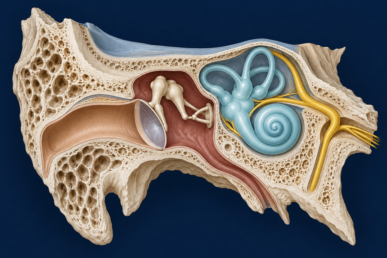 ILLUSTRATION · NOT CT / MRI
Stylised, simulated anatomy for orientation only; not to scale and not validated for identifying subtle signs. Animation is illustrative motion, not slice navigation or disease progression. Diagnostic evidence comes from the simulated reports.
Three boundaries to interrogate
Middle ear and ossicles
Review the attic, scutum, ossicular chain and routes into the mastoid. A report of soft tissue alone is not a tissue diagnosis.